The Biophysical World Inside a Jam-Packed Cell
Innovations in imaging and genetic engineering are coming together to probe the biophysics of cytoplasm inside living animals. The post The Biophysical World Inside a Jam-Packed Cell first appeared on Quanta Magazine
In recent years, the field of biophysics has witnessed a surge in innovation, driven by advancements in imaging technology and genetic engineering. These breakthroughs are now enabling scientists to probe the complex biophysical world inside the cytoplasm of living cells, offering unprecedented insights into cellular processes. This article delves into the intricate dance of molecules within the crowded cytoplasm, exploring how new tools are transforming our understanding of cellular biology.
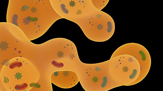
The cytoplasm, often depicted as a sparse environment in textbook diagrams, is actually a highly dynamic and congested space teeming with proteins, lipids, nucleic acids, and other cellular components. For decades, visualizing this intricate network has been a significant challenge. Traditional microscopy techniques have struggled to capture the full complexity of the cytoplasm due to the limited resolution and the sheer density of molecules. However, recent advancements in super-resolution microscopy and other imaging technologies have begun to change this landscape.
Super-resolution microscopy, a technique that allows for the visualization of cellular structures at a level far beyond the diffraction limit of conventional light microscopy, has proven particularly valuable. By using specialized fluorescent dyes or photoactivatable proteins, researchers can now image individual molecules within the cytoplasm with remarkable precision. This has revealed a previously hidden world of intricate molecular interactions and dynamic processes that were invisible to earlier methods.
In addition to imaging innovations, genetic engineering has also played a pivotal role in unraveling the biophysics of the cytoplasm. By tagging specific proteins or other molecules with fluorescent markers, scientists can track their movement and interactions within living cells. This approach, known as fluorescence resonance energy transfer (FRET), has provided critical insights into the spatial organization of cellular machinery and the mechanisms by which molecules navigate the crowded cytoplasm.
One of the most fascinating aspects of studying the cytoplasm is the way molecules navigate this dense environment. The cytoplasm is often likened to a "jam-packed" space, where molecules must constantly maneuver around one another to carry out their functions. Researchers have long wondered how these molecules find their way through this crowded landscape. Recent studies have shown that the cytoplasm is not a random, chaotic space but rather an organized and regulated environment.
Molecular motors, such as kinesin and dynein, play a crucial role in transporting cargo within the cytoplasm. These motor proteins move along microtubule tracks, carrying vesicles, organelles, and other cargo to their destinations. By visualizing these motors in action using advanced microscopy techniques, scientists have gained a better understanding of how cargo is transported and how this process is regulated.
Moreover, the cytoplasm is not static; it is a dynamic and evolving space that responds to cellular signals and environmental cues. For instance, during cell division, the cytoplasm undergoes dramatic reorganization to form the mitotic spindle, a structure essential for chromosome segregation. By studying these dynamic processes, researchers are uncovering the biophysical principles that govern cellular behavior.
The integration of imaging and genetic engineering has also opened new avenues for studying the biophysics of the cytoplasm in living animals. By combining these approaches, scientists can observe cellular processes in their natural context, without the need for tissue dissection or other invasive techniques. This has led to the discovery of novel mechanisms and pathways that were previously inaccessible to study.
One notable example is the use of genetically encoded fluorescent proteins, such as GFP (green fluorescent protein), to tag specific cellular components. By expressing these proteins in specific tissues or under certain conditions, researchers can visualize the spatial and temporal dynamics of cellular processes in living organisms. This has provided valuable insights into diseases and developmental processes, offering potential avenues for therapeutic interventions.
In conclusion, the biophysical world inside a jam-packed cell is a fascinating and complex realm that has long been shrouded in mystery. Innovations in imaging and genetic engineering are now enabling scientists to peer into this intricate network of molecules and uncover the biophysical principles that govern cellular function. As our understanding of the cytoplasm deepens, so too will our grasp of the fundamental processes that underlie life itself. The future of cellular biology looks brighter than ever, with these tools poised to unlock new frontiers of discovery.