Spectroscopic OCT plus AI detects high-risk plaque in coronary arteries
Artificial intelligence-based optical coherence tomography could improve long-term management of patients with coronary artery disease The post Spectroscopic OCT plus AI detects high-risk plaque in coronary arteries appeared first on Physics World .
In a breakthrough that could revolutionize the management of coronary artery disease, researchers in Korea have developed an AI-enhanced spectroscopic optical coherence tomography (S-OCT) system capable of detecting high-risk lipid-rich plaques in coronary arteries. This innovative approach combines the biochemical specificity of S-OCT with artificial intelligence (AI) to enable automated, composition-aware tissue characterization and lipid mapping. The technique offers interpretable and annotation-efficient lipid mapping, providing a scalable foundation for downstream assessment of lipid burden and clinically relevant plaque characterization, with potential utilization for automated risk stratification.
Lipid-rich plaques are critical in assessing a patient's risk of experiencing a heart attack. These fatty deposits adhere to the walls of blood vessels, and if they rupture, they can trigger adverse cardiovascular events. Currently, physicians rely on near-infrared spectroscopy and intravascular ultrasound (NIRS-IVUS) to quantitatively assess plaque lipid burden. While these methods are effective, they have limitations. Optical coherence tomography (OCT), another intravascular imaging modality used during catheter-based procedures, provides micrometre-resolution visualization of plaque structure. However, its diagnostic accuracy is often hindered by imaging artefacts and signals originating from non-lipid plaque components.
The new AI-enhanced S-OCT system addresses these challenges by integrating the biochemical specificity of S-OCT with AI. This combination enables automated, composition-aware tissue characterization and offers interpretable and annotation-efficient lipid mapping. The AI model, developed by researchers at the Korea Advanced Institute of Science and Technology (KAIST) and the Multimodal Imaging and Theranostic Lab of the Korea University Guro Hospital, utilizes existing OCT systems without requiring hardware modification.
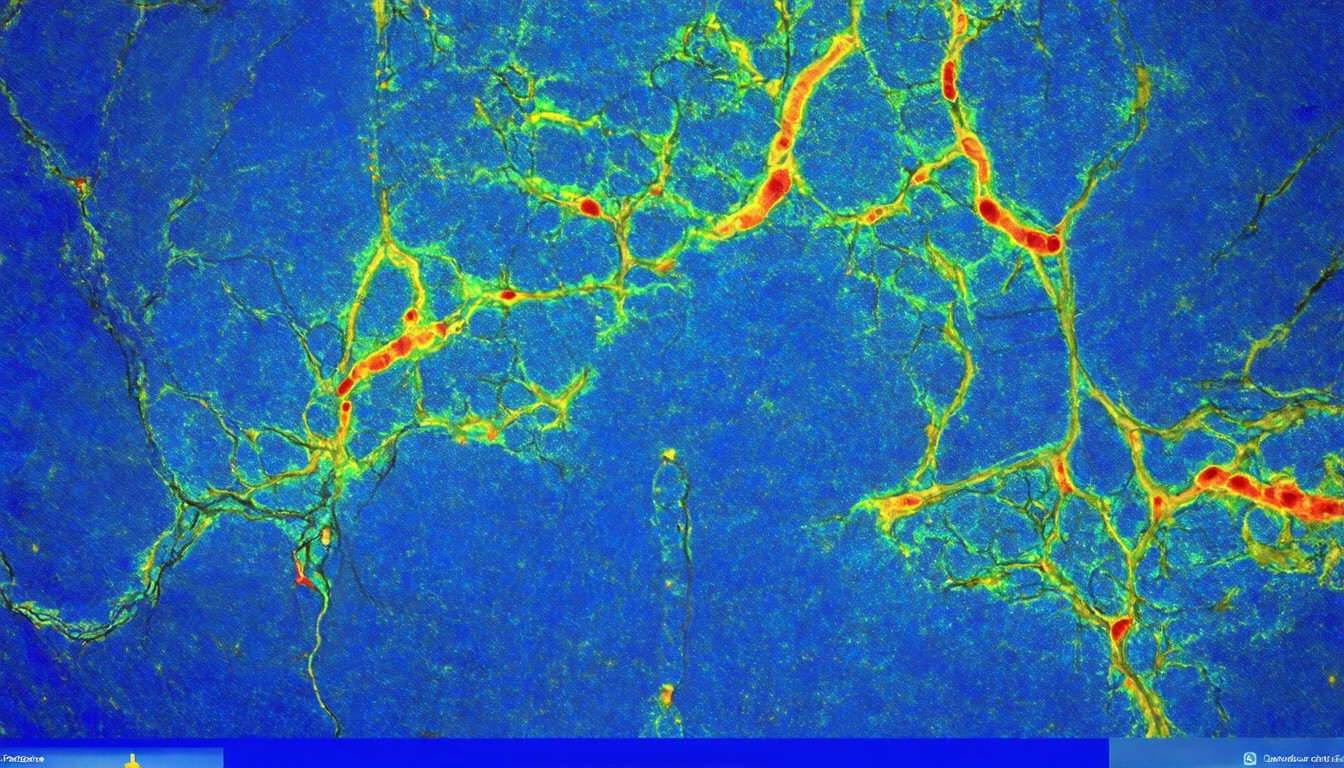
The researchers trained and validated the AI model using a dataset of intravascular OCT images and histopathology results. They found that the AI-identified lipid regions matched well with histopathology results, demonstrating the technique's accuracy and potential for clinical use. The AI-generated lipid maps overlay red areas indicating higher lipid likelihood, allowing physicians to identify areas of high-risk plaque more effectively.
This development has significant implications for the long-term management of patients with coronary artery disease. By enabling efficient lipid screening and spatial interpretation, the AI-enhanced S-OCT system provides a scalable foundation for downstream assessment of lipid burden and clinically relevant plaque characterization. With the potential for automated risk stratification, this technology could improve patient outcomes by enabling earlier and more accurate identification of high-risk patients, allowing for timely interventions and personalized treatment plans.
In conclusion, the integration of AI with spectroscopic OCT represents a promising advancement in the detection of high-risk plaques in coronary arteries. This innovative approach not only enhances diagnostic accuracy but also offers a more efficient and scalable method for lipid burden assessment. As the technology continues to evolve, it holds the potential to transform the management of coronary artery disease, ultimately improving patient outcomes and reducing the burden of cardiovascular diseases.